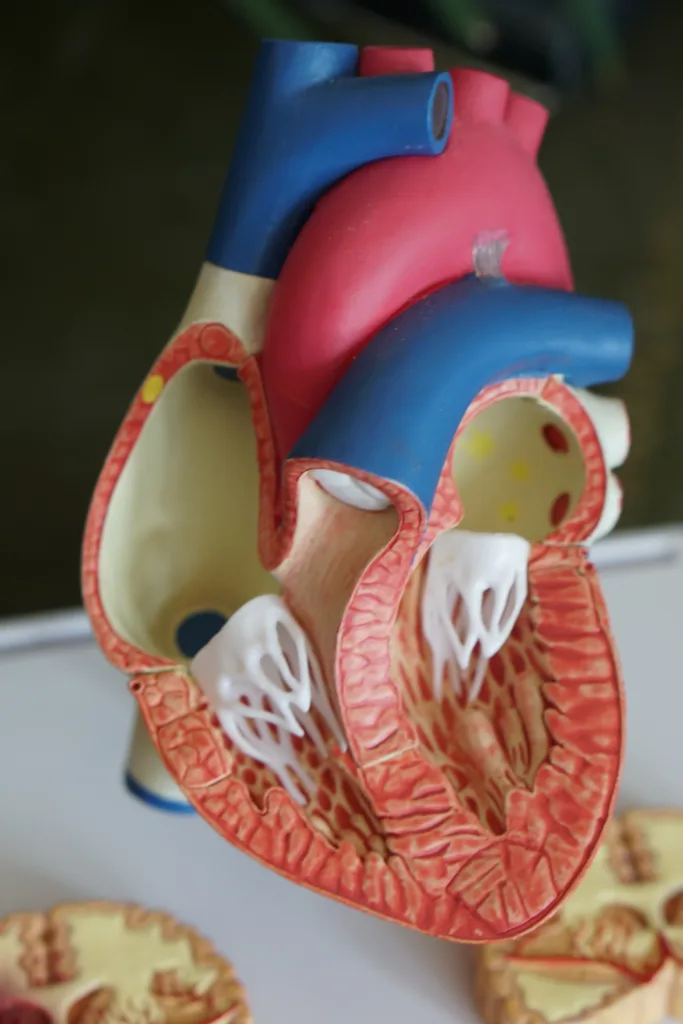
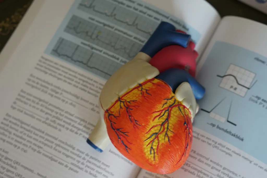
When it comes to understanding the functioning of the heart, the contraction of the ventricular walls plays a crucial role. The ventricles, which are the lower chambers of the heart, are responsible for pumping blood out of the heart and into the pulmonary arteries and the aorta.
During the contraction of the ventricular walls, a series of events take place that ensure the efficient flow of blood and prevent any backflow. One important component of this process is the presence of valves within the heart.
The tricuspid valve, located between the right atrium and right ventricle, is one such valve that plays a vital role in the overall functioning of the heart. It prevents the backflow of blood from the ventricles into the atria during the contraction phase.
As the ventricular walls contract, the pressure within the ventricles increases. This increase in pressure allows the blood to be propelled out of the heart and into the pulmonary arteries, which carry oxygen-depleted blood to the lungs, and the aorta, which carries oxygen-rich blood to the rest of the body.
The contraction of the ventricular walls is initiated by electrical impulses generated by the sinoatrial (SA) node. This node, also known as the natural pacemaker of the heart, sends an electrical signal through both atria, causing them to contract and push blood into the ventricles.
The electrical impulse then reaches the atrioventricular (AV) node, which is located between the atria and ventricles. The AV node continues to transmit the impulse, sending it down a specialized pathway known as the Bundle of His.
From the Bundle of His, the electrical signal branches into the right and left bundle branches, which further divide into smaller fibers called Purkinje fibers. These fibers extend throughout the ventricles, allowing the electrical impulse to reach every part of the ventricular walls.
As the electrical impulse reaches the Purkinje fibers, it triggers the contraction of the ventricular walls. This coordinated contraction of both ventricles ensures that blood is efficiently pumped out of the heart.
The contraction of ventricular walls is a highly coordinated process that involves the generation and transmission of electrical impulses through the heart. This process allows for the efficient pumping of blood and ensures that it flows in only one direction, preventing any backflow.
The contraction of the ventricular walls is a crucial step in the functioning of the heart. It is initiated by electrical impulses generated by the SA node and transmitted through the AV node, Bundle of His, bundle branches, and Purkinje fibers. This coordinated contraction allows for the efficient pumping of blood out of the heart and into the pulmonary arteries and aorta.
When The Ventricular Walls Contract What Happens?
When the ventricular walls contract, several important physiological changes occur in the heart. Here is a detailed explanation of what happens during ventricular contraction:
1. Increased pressure: As the ventricular walls contract, they exert force on the blood inside the chambers. This leads to an increase in pressure within the ventricles.
2. Ejection of blood: The increased pressure causes the semilunar valves (pulmonary valve and aortic valve) to open, allowing blood to be ejected from the ventricles. The right ventricle pumps blood into the pulmonary artery, which carries oxygen-depleted blood to the lungs. The left ventricle pumps blood into the aorta, which distributes oxygen-rich blood to the rest of the body.
3. Closure of atrioventricular valves: Simultaneously, the increased pressure within the ventricles causes the atrioventricular valves (tricuspid valve and mitral valve) to close. This prevents blood from flowing back into the atria and ensures that the blood is directed towards the arteries.
4. Ventricular relaxation: After the contraction, the ventricular walls relax, allowing the chambers to refill with blood. This phase is called diastole, and it prepares the heart for the next cycle of contraction and ejection.
When the ventricular walls contract, the pressure in the ventricles increases, leading to the ejection of blood into the pulmonary arteries and the aorta. At the same time, the atrioventricular valves close to prevent backflow of blood. This coordinated contraction and relaxation of the ventricles play a crucial role in pumping blood efficiently throughout the body.

When The Ventricular Walls Contract What Occurs To Prevent Backflow?
When the ventricular walls contract, several mechanisms work together to prevent backflow of blood. These mechanisms include:
1. Closure of the tricuspid valve: The tricuspid valve, located between the right atrium and right ventricle, closes tightly when the ventricular walls contract. This prevents the backflow of blood from the ventricle into the atrium.
2. Opening of the pulmonary valve: Simultaneously, the contraction of the right ventricle causes the pulmonary valve, situated between the right ventricle and the pulmonary artery, to open. This allows blood to be pumped out of the heart and into the pulmonary artery, which leads to the lungs.
3. Closure of the mitral valve: In the left side of the heart, the mitral valve, positioned between the left atrium and left ventricle, closes tightly during ventricular contraction. This prevents the backflow of blood from the left ventricle into the left atrium.
4. Opening of the aortic valve: As the left ventricle contracts, the aortic valve, situated between the left ventricle and the aorta, opens. This allows blood to be pumped out of the heart and into the aorta, which distributes oxygenated blood to the rest of the body.
When the ventricular walls contract, the tricuspid and mitral valves close tightly, while the pulmonary and aortic valves open. This coordinated action ensures that blood flows in only one direction, preventing any backflow and facilitating efficient circulation throughout the body.
When The Ventricular Walls Contract Enough To Eject Blood?
During the cardiac cycle, when the ventricular walls contract with sufficient force, blood is ejected from the ventricles into the pulmonary artery and aorta. This specific phase is known as systole. Systole can be further divided into two phases: isovolumetric contraction and ventricular ejection.
1. Isovolumetric Contraction: During this phase, the ventricles contract, but the volume of blood remains constant as both the atrioventricular (AV) valves and semilunar valves are closed. This closure prevents the backflow of blood into the atria and the outflow into the arteries. The ventricular pressure increases rapidly during isovolumetric contraction until it surpasses the pressure in the arteries.
2. Ventricular Ejection: Once the ventricular pressure exceeds the arterial pressure, the semilunar valves open, allowing blood to be ejected from the ventricles into the pulmonary artery and aorta. This ejection occurs due to the force generated by the contracting ventricular muscles. The blood is pushed into the respective arteries, ensuring proper circulation throughout the body.
During ventricular ejection, there are two main factors affecting the volume of blood ejected: stroke volume and ejection fraction.
– Stroke Volume: Stroke volume refers to the volume of blood ejected by each ventricle during one heartbeat. It is influenced by factors such as preload (volume of blood in the ventricles before contraction), afterload (resistance the ventricles must overcome to eject blood), and contractility (force generated by the ventricular muscles).
– Ejection Fraction: Ejection fraction is the percentage of blood ejected from the ventricle compared to the total volume of blood present in the ventricle. It is an important measure of cardiac function. A normal ejection fraction ranges from 50-70%. A decrease in ejection fraction may indicate a decrease in cardiac output and potential heart dysfunction.
During systole, the ventricular walls contract forcefully enough to generate pressure and eject blood into the pulmonary artery and aorta. This process involves the phases of isovolumetric contraction and ventricular ejection. Factors such as stroke volume and ejection fraction play a crucial role in determining the efficiency of blood ejection from the ventricles.
What Causes The Ventricles To Contract?
The ventricles contract due to the electrical signals sent by the sinoatrial (SA) node, atrioventricular (AV) node, Bundle of His, bundle branches, and Purkinje fibers. Here is a step-by-step explanation of how this process occurs:
1. The SA node, located in the right atrium, generates electrical impulses.
2. The SA node sends these impulses through both atria, causing them to contract.
3. The impulses reach the AV node, which is located between the atria and ventricles.
4. The AV node delays the impulse slightly to allow the atria to fully contract and for blood to flow into the ventricles.
5. After the delay, the AV node continues to send the impulse down the Bundle of His.
6. The Bundle of His divides into right and left bundle branches, which extend into the ventricles.
7. The impulse travels through the bundle branches, stimulating the Purkinje fibers.
8. The Purkinje fibers distribute the impulse throughout the ventricles, causing them to contract simultaneously.
9. The coordinated contraction of the ventricles forces blood out of the heart and into the arteries.
The electrical signals generated by the SA node and transmitted through the AV node, Bundle of His, bundle branches, and Purkinje fibers are responsible for causing both ventricles to contract and pump blood out of the heart.

Conclusion
The ventricular walls play a crucial role in the pumping action of the heart. As the ventricles contract, the pressure within them increases, allowing for the efficient ejection of blood into the pulmonary arteries and aorta. This process is facilitated by the opening and closing of valves that prevent backflow of blood into the ventricles.
The tricuspid valve, located between the right atrium and right ventricle, ensures that blood flows in only one direction. This valve, along with other valves in the heart, ensures the proper circulation of blood throughout the body.
The contraction of the ventricles is coordinated by the electrical impulses generated by the sinoatrial (SA) node. This impulse travels through both atria, causing them to contract, and then reaches the atrioventricular (AV) node. From there, the impulse passes through specialized fibers known as the Bundle of His, right and left bundle branches, and eventually reaches the Purkinje fibers within the ventricles. This coordinated electrical activity ensures that both ventricles contract simultaneously, allowing for efficient pumping of blood.
Understanding the intricate workings of the ventricular walls and their role in the heart’s pumping action is essential for comprehending the overall function of the cardiovascular system. By maintaining proper pressure and direction of blood flow, the ventricular walls contribute to the overall health and vitality of the body.
