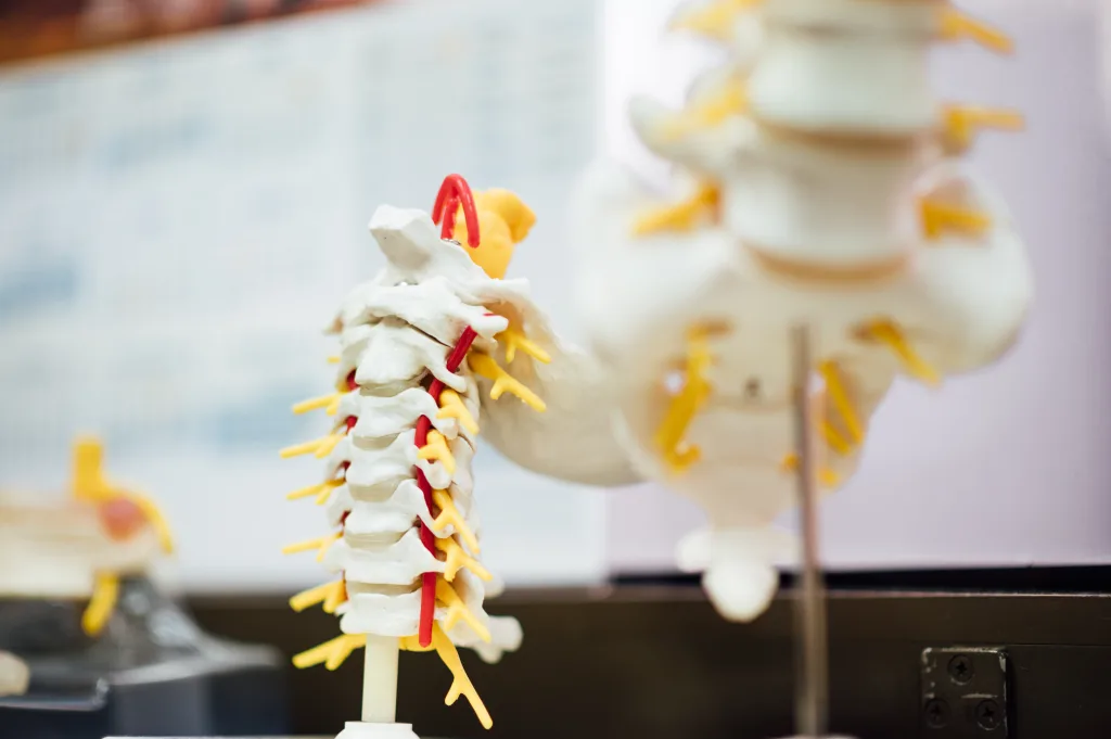
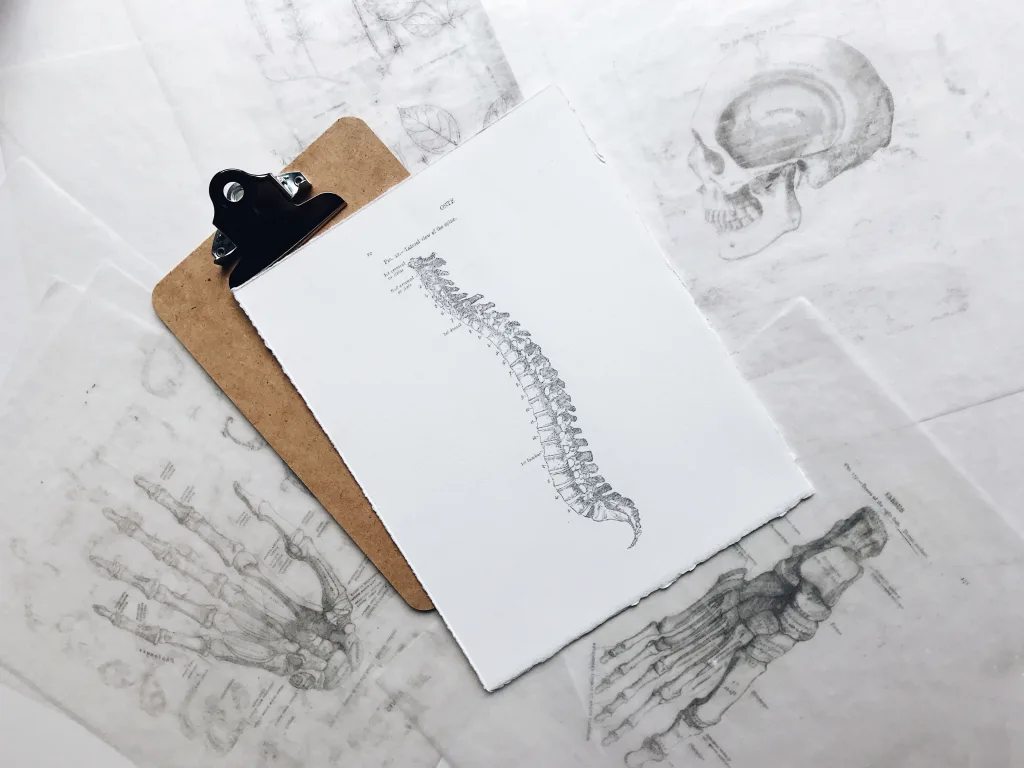
The thecal sac, also known as the dural sac, is a protective membrane that surrounds the spinal cord and cauda equina. This membranous sheath is made up of the dura mater, one of the layers of the spinal meninges. Its primary function is to provide a barrier and support for the delicate spinal cord and its associated nerves.
The thecal sac contains cerebrospinal fluid (CSF) in which the spinal cord “floats”. This fluid serves as a cushion, protecting the spinal cord from injury and providing nutrients to its tissues. It also helps maintain the optimal environment for the proper functioning of the spinal cord.
In adults, the dural sac terminates caudally at the second sacral segment (S2). This means that it extends down to the level of the second bone in the sacrum, which is a triangular bone located below the lumbar spine. The termination of the spinal cord itself, known as the conus medullaris, is slightly higher and typically ends at the level of the first and second lumbar vertebrae (L1-2).
It is important to note that the termination point of the dural sac and the spinal cord can vary depending on the individual. In newborns, the dural sac ends at the third sacral segment (S3), and the conus medullaris terminates at the third lumbar vertebra (L3). These differences are due to the growth and development of the spinal column as an individual ages.
At the base of the skull, the dural sac passes through a large opening called the foramen magnum. As it enters the cranial cavity, it becomes continuous with the layer of dura mater that surrounds and protects the brain. This connection allows for the exchange of CSF between the spinal cord and the brain.
At the lower end of the vertebral canal, within the sacrum, the dural sac tapers down to a point. This occurs at the level of the second sacral segment (S2). The narrowing of the dural sac in this region is a natural anatomical feature and does not typically cause any issues.
The thecal sac, or dural sac, is a protective membrane that surrounds the spinal cord and cauda equina. It contains cerebrospinal fluid and provides support and cushioning for the spinal cord. In adults, the dural sac terminates at the second sacral segment (S2), while the spinal cord itself ends slightly higher at the level of the first and second lumbar vertebrae (L1-2). Understanding the anatomy of the thecal sac is important for diagnosing and treating spinal conditions and ensuring the proper functioning of the spinal cord.
Where Does The Thecal Sac Start?
The thecal sac starts at the base of the skull, specifically at the foramen magnum, which is the large opening at the bottom of the skull where the spinal cord exits. From there, it extends down the entire length of the spinal column, surrounding and protecting the spinal cord and cauda equina. The thecal sac is continuous with the dura mater, which is the outermost layer of the spinal cord and brain. It encloses the cerebrospinal fluid that cushions and supports the spinal cord, allowing it to float within the thecal sac.

Where Is The Thecal Sac Located?
The thecal sac is located within the spinal column and surrounds the spinal cord. More specifically, it is situated within the vertebral canal, which is formed by the stacked vertebrae of the spine. The thecal sac runs down the length of the spinal canal and contains the spinal cord, spinal nerve roots, and cerebrospinal fluid (CSF). It acts as a protective covering for the spinal cord, shielding it from potential damage.
Where Does Dural Sac End?
The dural sac is a protective covering that surrounds the spinal cord and extends downwards. In an adult, it terminates at the level of the second sacral vertebra, which is referred to as S2. This means that the dural sac covers the spinal cord up until this point.
It is important to note that the termination of the dural sac may vary depending on the age of the individual. In newborns, the dural sac ends at the third sacral vertebra, which is called S3. As the individual grows and develops, the dural sac gradually extends further down the spine.
Where Does The Dural Sac Start And End?
The dural sac, a protective layer surrounding the spinal cord, begins at the base of the skull and extends down to the sacrum, which is the triangular bone located at the base of the spine. More specifically, the dural sac starts at the level of the foramen magnum, which is the large opening at the base of the skull through which the spinal cord passes. From there, it continues downwards through the vertebral canal, which is the hollow space formed by the vertebral bones.
As the dural sac descends, it gradually narrows and tapers down to a point. This narrowing occurs at the level of the second sacral segment, which is the second vertebra in the sacrum. At this point, the dural sac comes to an end.
The dural sac begins at the base of the skull, passing through the foramen magnum, and continues down through the vertebral canal until it tapers down to a point at the level of the second sacral segment in the sacrum.

Conclusion
The thecal sac is a crucial component of the spinal cord and cauda equina, providing a protective sheath of dura mater. It surrounds and contains the cerebrospinal fluid, allowing the spinal cord to float within it. The thecal sac acts as the outer covering of the spinal cord, shielding it from external pressures and potential damage.
The termination point of the thecal sac varies between adults and newborns. In adults, it ends at the S2 level, while in newborns, it ends at S3. The spinal cord itself terminates at the L1-2 level in adults, known as the conus medullaris, and at the L3 level in newborns.
At the base of the skull, the thecal sac passes through the foramen magnum and becomes continuous with the layer of dura mater that surrounds the brain. At the lower end, within the vertebral canal of the sacrum, the thecal sac narrows down to a point, reaching the second sacral segment.
Understanding the anatomy and function of the thecal sac is essential for diagnosing and treating conditions that may affect it. Bone spurs or other abnormalities can put pressure on the front part of the thecal sac, potentially impacting the spinal cord and causing various symptoms. By recognizing these issues and providing appropriate care, healthcare professionals can help ensure the health and well-being of patients.
