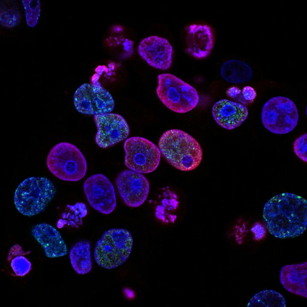
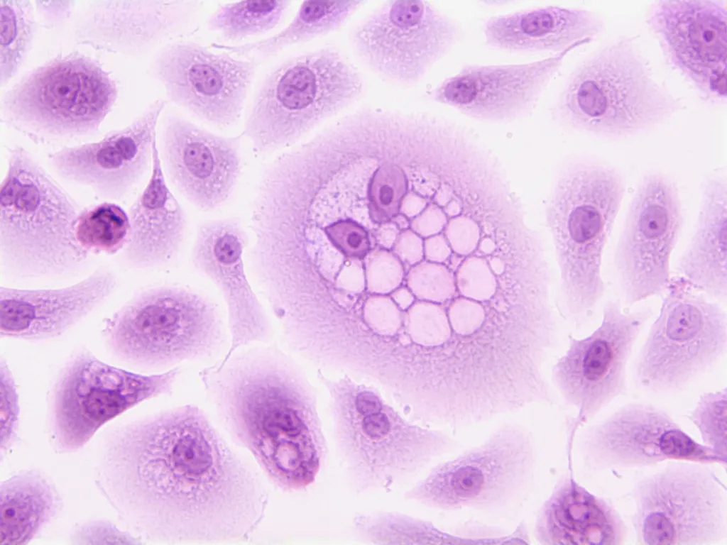
The highlighted structure in the image is the nuclei. Nuclei are essential components of cells and play a vital role in maintaining the cell’s genetic information. They are often referred to as the “control center” of the cell because they contain the DNA, which carries the instructions for the cell’s functions and activities.
Nuclei are found in almost all eukaryotic cells, including those in plants, animals, and fungi. They are typically spherical or oval-shaped and are surrounded by a double membrane called the nuclear envelope. The nuclear envelope separates the contents of the nucleus from the rest of the cell, ensuring that the DNA is protected and regulated.
Within the nucleus, there are various structures and components that contribute to its functions. One of the most prominent structures is the nucleolus, which is involved in the production of ribosomes, the cellular machinery responsible for protein synthesis. The nucleolus appears as a dense, dark spot within the nucleus.
The highlighted nuclei in the image are most likely from a tissue sample, as individual cells are not usually visible in isolation. The tissue appears to be composed of stratified squamous epithelium, a type of epithelial tissue that is found in areas subjected to mechanical stress, such as the skin, oral cavity, and esophagus. The stratified squamous epithelium is characterized by multiple layers of cells, with the outermost layer being flattened and squamous in shape.
In the image, the nuclei within the stratified squamous epithelium are visible as dark, round structures. Each cell within the tissue has a single nucleus, which contains the genetic material necessary for cellular functions and replication.
It is worth noting that the presence of multiple layers of cells in the stratified squamous epithelium allows for increased protection against mechanical stress and damage. The arrangement of the cells also allows for the shedding of dead cells from the surface, ensuring the maintenance of a healthy and functional epithelial barrier.
The highlighted structure in the image is the nuclei, which are essential components of cells and contain the genetic material necessary for cellular functions. The nuclei are visible within a tissue sample of stratified squamous epithelium, a type of epithelial tissue found in areas subjected to mechanical stress. The stratified arrangement of the cells in this tissue provides increased protection and allows for the shedding of dead cells from the surface.
What Are The Highlighted Structures?
The highlighted structures in the image appear to be nuclei. Nuclei are the central organelles found within cells, which contain the cell’s genetic material and play a crucial role in regulating cellular activities. In this image, the nuclei are likely from a specific type of tissue or organ, although without further context, it is difficult to determine their exact origin.
Additionally, the highlighted epithelium in the image appears to be stratified squamous epithelium. Stratified squamous epithelium is a type of tissue that consists of multiple layers of flat cells. It is commonly found in areas of the body that experience mechanical stress, such as the skin, oral cavity, esophagus, and vagina. This type of epithelium provides protection against abrasion and serves as a barrier against pathogens and other harmful substances.
The highlighted structures in the image are nuclei and the highlighted epithelium is stratified squamous epithelium.

What Structure Connects The Highlighted Muscle Cell To One Another?
The structure that connects the highlighted muscle cell to one another is called intercalated discs. Intercalated discs are specialized cell junctions that play a crucial role in maintaining the structural integrity and functional coordination of cardiac muscle tissue. These discs consist of two main types of junctions: anchoring junctions and gap junctions.
1. Anchoring junctions: These junctions, also known as desmosomes, are responsible for providing strong adhesion between adjacent cardiomyocytes. They prevent the cells from separating during contraction and relaxation of the heart muscle. Anchoring junctions consist of proteins called cadherins, which link the cytoskeletons of neighboring cells together, creating a mechanical connection.
2. Gap junctions: Gap junctions facilitate direct communication between adjacent cardiomyocytes. They are formed by protein channels called connexons, which allow the exchange of ions, small molecules, and electrical impulses between cells. This communication is crucial for coordinating the contraction and relaxation of the heart muscle, ensuring efficient pumping of blood.
Intercalated discs in cardiac muscle tissue connect muscle cells together through anchoring junctions to maintain structural integrity and through gap junctions to facilitate communication and coordination between cells.
Which Epithelial Type Is Highlighted Simple Columnar Epithelium?
The highlighted tissue is simple columnar epithelium. Simple columnar epithelium is a type of epithelial tissue that is composed of a single layer of tall, column-shaped cells. These cells are closely packed together and have elongated nuclei. This type of epithelium is typically found lining the digestive tract, where it plays a role in absorption and secretion. It is also found in other areas of the body, such as the respiratory tract and the reproductive system. Simple columnar epithelium is characterized by its tall, narrow shape and the presence of microvilli on the apical surface, which increase the surface area for absorption. It is also involved in the secretion of mucus and enzymes. Simple columnar epithelium can be found in the small intestine, where it lines the surface of the intestinal villi and aids in the absorption of nutrients. In the stomach, it forms gastric pits and secretes gastric juices. In the reproductive system, it lines the fallopian tubes and plays a role in the transport of eggs. simple columnar epithelium is an important tissue type that is specialized for absorption and secretion in various parts of the body.
What Structures Are Found On The Apical Surface Of The Highlighted Epithelium?
On the apical surfaces of epithelial cells, several specialized structures can be found. These structures include:
1. Cilia: These are hair-like projections that extend from the apical surface of the cell. They are composed of microtubules and are involved in various functions such as the movement of mucus and other substances along the surface of the epithelium.
2. Microplicae: These are tiny folds or ridges on the apical surface of the cell. They increase the surface area of the cell, allowing for better absorption and secretion of substances.
3. Microvilli: These are small finger-like projections that cover the apical surface of the cell. They also increase the surface area of the cell, aiding in absorption and secretion processes. Microvilli are particularly abundant in cells involved in absorption, such as those lining the small intestine.
4. Stereocilia: These are long, non-motile microvilli found in certain specialized epithelial cells, such as the epididymis in the male reproductive system and the hair cells of the inner ear. They play a role in sensory processes and are involved in the detection of sound and movement.
The highlighted epithelium displays various structures on its apical surface, including cilia, microplicae, microvilli, and stereocilia. These structures serve different functions, such as movement, absorption, secretion, and sensory detection.

Conclusion
The highlighted structure in the image is stratified squamous epithelium. This type of epithelium is characterized by multiple layers of flat, scale-like cells. It is found in areas of the body that experience mechanical stress and abrasion, such as the skin, oral cavity, esophagus, and vagina. Stratified squamous epithelium provides protection against physical damage, pathogens, and dehydration. It also plays a role in the secretion and absorption of certain substances. The cells of stratified squamous epithelium are tightly packed together and adhere to one another through specialized cell junctions. These junctions, known as intercalated discs, contain both anchoring junctions and gap junctions. Anchoring junctions help to hold the cells together, while gap junctions allow for the direct communication and exchange of ions and molecules between adjacent cells. This structure allows for the coordinated contraction and electrical coupling of neighboring cells, which is essential for the functioning of tissues and organs.
