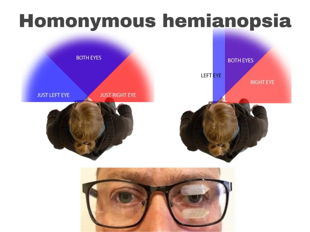
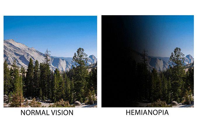
Hemianopia is a condition that is caused by damage to the brain, which can be the result of a stroke, trauma or tumour. This damage affects the area of the brain that is responsible for processing and interpreting visual information from the eyes. As a result, the person who has hemianopia experiences a loss of vision in one half of their visual field.
The extent of the field loss can vary depending on the location and severity of the brain damage. In some cases, the person may only lose a small portion of their visual field, while in other cases they may lose almost the entire half of their visual field.
Ischemic and hemorrhagic strokes are the most common causes of hemianopia in adults. Ischemic strokes occur when a blood clot blocks a blood vessel in the brain, while hemorrhagic strokes occur when a blood vessel in the brain ruptures. Both types of stroke can cause damage to the brain that leads to hemianopia.
In children, hemianopia is less common than in adults, but when it does occur, it is often caused by a stroke. Studies have shown that approximately 25% of children with hemianopia have had a stroke.
Homonymous superior quadrantanopia is a specific type of hemianopia that is caused by damage to the contralateral inferior parts of the posterior visual pathway. This can include the inferior optic radiation (temporal Meyer loop), or the inferior part of the occipital visual cortex beow the calcarine fissure.
Hemianopia is a condition that is caused by damage to the brain, which can result from a stroke, trauma or tumour. The extent of the field loss can vary depending on the location and severity of the brain damage. It is important to seek medical attention if you experience any changes to your vision, as early diagnosis and treatment can help to prevent further damage to the brain.
Causes of Hemianopia
Hemianopia is a visual disorder that results from damage to the brain. This damage can be caused by various factors, including a stroke, a head injury, a tumor, or any other condition that affects the brain. The extent of the visual field loss depends on the location of the damage in the brain.
A stroke is one of the most common caues of hemianopia. A stroke occurs when the blood supply to the brain is interrupted, causing brain cells to die. Depending on where the stroke occurs, it can cause different types of visual field loss. For example, a stroke in the occipital lobe of the brain, which is responsible for processing visual information, can cause homonymous hemianopia, where the loss of vision affects the same side of both eyes.
Trauma to the head can also cause hemianopia. This can occur from a direct blow to the head, such as in a car accident, or from a concussion. Hemianopia caused by trauma can be temporary or permanent, depending on the severity of the injury.
A brain tumor can also cause hemianopia. Tumors can grow in different parts of the brain and can cause damage to the visual processing areas of the brain. This can result in visual field loss that can be either temporary or permanent, depending on the size and location of the tumor.
Hemianopia is caused by damage to the brain, which can result from various factors such as stroke, head injury, or a brain tumor. The extent of visual field loss depends on the location and severity of the damage to the brain.

Effects of Hemianopia Caused by Stroke
Hemianopia is a visual field loss that can occur following a stroke. There are two main types of stroke that can cause hemianopia: ischemic stroke and hemorrhagic stroke.
Ischemic stroke occurs when a blood clot blocks a blood vessel in the brain, leading to a lack of oxygen and nutrients to the affected area. This type of stroke is the most common cause of hemianopia, accounting for up to 80% of cases.
Hemorrhagic stroke, on the other hand, occurs when a blood vessel in the brain ruptures and causes bleeding into the surrounding tissue. While less common than ischemic stroke, hemorrhagic stroke can also lead to hemianopia.
It’s worth noting that hemianopia resulting from stroke is not limited to adults. In fact, approximately 25% of cases in children are also caused by stroke.
Both ischemic and hemorrhagic strokes can cause hemianopia, with ischemic stroke being the most common cause.
What is Hemianopia?
Hemianopia is a visual impairment that affects half of your visual field. This condition is not caused by any problem with your eyes. Instead, it occurs when there is damage to the brain, most commonly after a stroke or brain injury.
Individuals with hemianopia may have difficulty seeing objects, people, or surroundings on one side of ther visual field. The extent of the visual impairment depends on the severity and location of the brain damage. Some people may experience only a partial loss of vision, while others may have a complete loss of sight in one half of their visual field.
Hemianopia can greatly impact an individual’s daily life, making it difficult to perform certain tasks such as driving, reading, or walking safely. It can also affect their orientation and spatial awareness, leading to a greater risk of falls and accidents.
In some cases, rehabilitation therapy such as visual field training or compensatory strategies can help individuals with hemianopia to improve their visual function and adapt to their condition. However, it is important to consult with a qualified healthcare professional for proper diagnosis and treatment.
Hemianopia is a visual impairment that affects half of the visual field, caused by brain damage after a stroke or injury. It can greatly impact an individual’s daily life, but rehabilitation therapy may help improve visual function and adaptation.
Causes of Superior Hemianopia
Superior hemianopia is a visual impairment that affects the upper half of the visual field of one or both eyes. This condition is caused by damage to the posterior visual pathway, specifically to the inferior parts of the visual cortex or the optic radiation.
The visual cortex is the part of the brain that processes visual information received from the eyes. It is located in the occipital lobe, at the back of the brain. The optic radiation is a group of nerve fibers that connects the visual cortex to the thalamus, which relays sensory information to other parts of the brain.
Damage to the inferior parts of the visual cortex or the optic radiation can result in superior hemianopia, as these areas are responsible for processing visual information from the lower half of the visual field. The most common causes of superior hemianopia include stroke, head trauma, tumors, and neurodegenerative diseases.
Stroke is the most common cause of superior hemianopia, accounting for about 70% of cases. In stroke, the blood flow to the brain is disrupted, leading to brain damage. The occipital lobe is particularly susceptible to stroke, as it is supplied by small, delicate blood vessels.
Head trauma, such as a blow to the head, can also cause superior hemianopia. This is because the impact can damage the optic radiation or the visual cortex.
Tumors and neurodegenerative diseases, such as Alzheimer’s disease and Parkinson’s disease, can also cause superior hemianopia. Tumors can compress the optic radiation or the visual cortex, whle neurodegenerative diseases can cause gradual damage to these areas over time.
Superior hemianopia is caused by damage to the inferior parts of the visual cortex or the optic radiation, usually as a result of stroke, head trauma, tumors, or neurodegenerative diseases.
The Brain Region Responsible for Homonymous Hemianopia
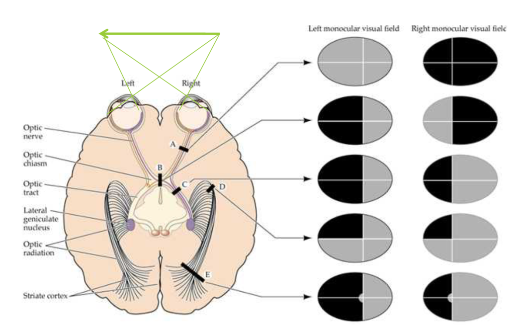
Homonymous hemianopia is a visual field defect that affects both eyes and results in loss of vision in half of the visual field, either the right or left side. This condition is caused by lesions in the retrochiasmal visual pathways, which are located in the posterior part of the brain.
The retrochiasmal visual pathways consist of several structures, including the optic tract, the lateral geniculate nucleus, the optic radiations, and the cerebral visual (occipital) cortex.
The optic tract is a bundle of nerve fibers that carries visual information from the eyes to the brain. It travels from the optic chiasm, where the optic nerves from each eye cross, to the lateral geniculate nucleus in the thalamus.
The lateral geniculate nucleus is a relay center in the thalamus that receives visual information from the optic tract and sends it to the visual cortex.
The optic radiations are a group of nerve fibers that connect the lateral geniculate nucleus to the visual cortex. They pass through several structures, including the internal capsule, which is a band of white matter in the brain that connects the cortex to oher parts of the brain.
The cerebral visual cortex is located in the occipital lobe of the brain and is responsible for processing visual information. It is divided into several areas, each with a specific function, such as detecting motion, color, and shape.
Lesions in any of these structures can cause homonymous hemianopia. For example, a stroke that affects the optic radiations on the left side of the brain can result in loss of vision in the right visual field of both eyes.
Homonymous hemianopia is caused by lesions in the retrochiasmal visual pathways, which include the optic tract, the lateral geniculate nucleus, the optic radiations, and the cerebral visual cortex.

Effects of Artery Occlusion on Homonymous Hemianopia
The occlusion of the middle cerebral artery (MCA) is known to cause homonymous hemianopia. This condition is characterized by the loss of vision in the contralateral visual field of both eyes. In other words, if the MCA is occluded on the left side of the brain, the patient will experience a loss of vision in the riht visual field of both eyes. This occurs because the MCA supplies blood to the primary visual cortex, which is responsible for processing visual information. When this area of the brain is damaged due to an MCA occlusion, it results in homonymous hemianopia. It is important to note that other symptoms may accompany an MCA occlusion, including paralysis and sensory loss of the contralateral face and upper limb, global aphasia if the dominant side is involved, and spatial neglect if the nondominant side is involved.
The Location of Hemianopia
Hemianopia, also knwn as hemianopsia, is a visual disorder that affects the same halves of the visual field of both eyes. This condition can occur as a result of cerebrovascular injury or tumor in the brain, which disrupts the normal functioning of the visual pathways. The damage can occur anywhere along the visual pathway, including the optic nerve, optic chiasm, or optic tract.
The optic nerve is responsible for transmitting visual information from the retina to the brain. If the optic nerve is damaged, it can lead to hemianopia in the corresponding visual field of both eyes.
Similarly, the optic chiasm is a point where the optic nerves from both eyes meet and cross over. If the optic chiasm is affected, it can disrupt the flow of visual information to the brain, resulting in hemianopia.
Lastly, the optic tract carries visual information from the optic chiasm to the visual cortex in the brain. Any damage to the optic tract can also cause hemianopia in the corresponding visual field of both eyes.
Hemianopia can occur at any point along the visual pathway, including the optic nerve, optic chiasm, or optic tract, as a result of cerebrovascular injury or tumor in the brain.
Vision Loss Caused by Stroke
Vision loss caused by a stroke is known as an eye stroke or anterior ischemic optic neuropathy. This condition occurs when there is a lack of sufficient blood flow to the tissues located in the front part of the optic nerve. The optic nerve is responsible for transmitting visual information from the eyes to the brain, and any damage or disruption to this nerve can result in vision loss.
Eye strokes are a type of ischemic stroke, which means they occur when blood flow to a specific part of the brain is blocked. In the case of an eye stroke, the blockage occurs in the blood vessels that supply the optic nerve with oxygen and nutrients. This lack of blood flow can caue the cells in the optic nerve to become damaged or die, leading to vision loss.
There are several risk factors that can increase the likelihood of experiencing an eye stroke, including high blood pressure, diabetes, smoking, and high cholesterol. Additionally, certain medical conditions such as giant cell arteritis and lupus can also increase the risk of an eye stroke.
Symptoms of an eye stroke can include sudden vision loss, blurred vision, or a visual field defect (a portion of the visual field is missing). Treatment for an eye stroke typically involves addressing the underlying cause, such as managing high blood pressure or diabetes, and may also include medications to improve blood flow to the optic nerve. In some cases, vision may improve over time, but in others, it may be permanent.
If you experience sudden vision loss or any other visual symptoms, it is important to seek medical attention immediately. Early treatment can help prevent further damage to the optic nerve and improve the chances of recovering some or all of your vision.
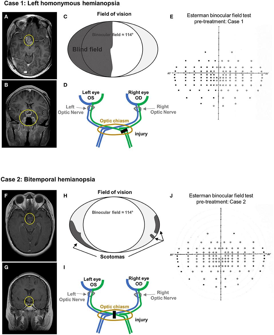
Causes of Left Homonymous Hemianopia
Homonymous hemianopia (HH) is a condition that results in the loss of half of the visual field in both eyes. Left homonymous hemianopia refers specifically to the loss of vision in the left half of both eyes.
The most common cause of left homonymous hemianopia is a stroke. Strokes occur when blood flow to the brain is disrupted, which can damage brain tissue and lead to a variety of neurological symptoms. If the stroke occurs in the right side of the brain, it can result in left homonymous hemianopia.
Other potential causes of left homonymous hemianopia include:
– Trauma: A blow to the head or other injury to the brain can cause damage that leads to visual field loss.
– Tumors: Brain tumors can grow and press on the optic nerve or other parts of the visual system, which can cause visual field loss.
– Infections: Certain infections, such as encephalitis, can cause inflammation in the brain that leads to visual symptoms.
– Other neurological conditions: Conditions such as multiple sclerosis or Alzheimer’s disease can also cause visual field loss.
In many cases, the location and characteristics of the visual field loss can help determine the underlying cause of left homonymous hemianopia. For example, stroke-related HH may be accompanied by other neurological symptoms such as weakness or numbness on the right side of the body, whie trauma-related HH may be more localized to the visual system.
If you or a loved one experiences sudden vision loss, it is important to seek medical attention right away. A comprehensive evaluation can help determine the underlying cause of the condition and guide appropriate treatment.

Source: frontiersin.org
Naming Hemianopsia
Hemianopsia is a medical condition that refers to the loss of vision or blindness in half of the visual field. It can affect one or both eyes, depending on the cause and severity of the condition. There are two main types of hemianopsia, which are classified based on the area of the visual field that is affected:
1. Binasal hemianopsia: This type of hemianopsia is characterized by the loss of vision in the areas surrounding the nose. This means that the person is unable to see objects or images that are located on the side of teir nose. Binasal hemianopsia is often caused by damage to the optic chiasm, which is the area where the optic nerves from both eyes cross over.
2. Bitemporal hemianopsia: This type of hemianopsia is characterized by the loss of vision in the areas closest to the temples. This means that the person is unable to see objects or images that are located on the side of their temples. Bitemporal hemianopsia is often caused by damage to the optic nerves, which are responsible for transmitting visual information from the eyes to the brain.
Hemianopsia is the medical condition that refers to the loss of vision or blindness in half of the visual field. It is named based on the area of the visual field that is affected, which can be either binasal or bitemporal.
The Effects of Diabetes on Hemianopia
Diabetes can cause hemianopia, which is a visual field defect that affects one half of the visual field in both eyes. This condition is usually caused by damage to the optic nerve or the visual cortex in the brain.
In patients with diabetes, high blood sugar levels can lead to a condition called nonketotic hyperglycemia, which can cause various neurologic symptoms, including hemianopia. This condition is more common in elderly patients with type 2 diabetes who have poor glycemic control.
Hyperglycemia can cause damage to the blood vessels in the retina, leading to a condition called diabetic retinopathy, which is a leading cause of blindness in diabetic patients. In some cases, diabetic retinopathy can progress to involve the optic nerve and lead to hemianopia.
In addition to hemianopia, patients with nonketotic hyperglycemia may present with other neurologic symptoms, such as confusion, seizures, and hemiparesis. These symptoms can be reversible with aggressive glycemic control and adequate hydration.
If you have diabetes and are experiencing any neurologic symptoms, it is important to seek medical attention immediately. Your healthcare provider can perform a comprehensive evaluation to determine the underlying cause of your symptoms and recommend appopriate treatment.
Causes of Partial Loss of Vision in One Eye
Partial loss of vision in one eye, also known as unilateral vision loss, can be caused by various factors. The most common cause is reduced blood flow to the eye, whih can be due to plaque buildup in the carotid arteries in the neck. Plaque is a fatty substance that accumulates on the walls of blood vessels, causing them to narrow and restrict blood flow. This can lead to a condition called amaurosis fugax, which is temporary vision loss in one eye.
Other possible causes of partial vision loss in one eye include:
– Optic neuritis: inflammation of the optic nerve, which can cause blurry vision or blindness in one eye.
– Macular degeneration: a condition that affects the central part of the retina, causing loss of vision in the center of the visual field.
– Retinal detachment: when the retina becomes detached from the back of the eye, causing vision loss in one eye.
– Glaucoma: a group of eye diseases that damage the optic nerve and can cause vision loss in one or both eyes.
It is important to seek medical attention if you experience any sudden or gradual vision loss in one eye, as it may be a symptom of a serious underlying condition. Your doctor will perform a thorough examination and may recommend further tests or treatments depending on the cause of your vision loss.
The Most Common Cause of Bitemporal Hemianopia
Bitemporal hemianopia, a condition where a person loses their peripheral vision from both eyes, is primarily caused by damage to the optic chiasm. The optic chiasm is a crucial point where the optic nerves from both eyes intersect, and any damage to this area can lead to a loss of vision.
The most common causs of bitemporal hemianopia are tumors and aneurysms. Tumors can grow in the pituitary gland, which is located near the optic chiasm, and compress the chiasm, leading to vision loss. Similarly, aneurysms in the arteries that supply blood to the brain can also press on the optic chiasm, causing damage.
Other less common causes of bitemporal hemianopia include inflammatory and ischemic diseases. Inflammatory diseases like sarcoidosis and multiple sclerosis can cause inflammation in the optic chiasm, leading to vision loss. Ischemic diseases like stroke and arteriosclerosis can also cause damage to the optic chiasm by limiting blood flow to the area.
The most common cause of bitemporal hemianopia is damage to the optic chiasm, which can occur from tumors, aneurysms, and less commonly, inflammatory and ischemic diseases.

Source: allaboutvision.com
Partial Hemianopia: Overview and Causes
Partial hemianopia is a visual impairment that occurs when a person loses less than half of ther field of vision. It can be caused by damage to the optic nerve, brain injury, or other medical conditions. A person with partial hemianopia may experience difficulty seeing objects on one side of their visual field, which can affect their ability to navigate their environment, read, and perform other daily tasks.
Quadrantanopia is a type of partial hemianopia where a person loses a quarter of their vision in both eyes, typically in one of the four quadrants of their visual field. This can also be caused by brain injury or medical conditions affecting the optic nerve.
Complete hemianopia, on the other hand, is when a person loses half of their field of vision. This can be caused by a stroke, brain injury, or other medical conditions. People with complete hemianopia may experience significant visual impairment and may require special accommodations to perform daily tasks.
It’s important for individuals experiencing any type of visual impairment to seek medical attention to determine the underlying cause and receive appropriate treatment. Depending on the severity of the impairment, accommodations such as visual aids or therapy may be recommended by healthcare professionals.
The Relationship Between Hemianopia and Blindsight
Hemianopia and blindsight are related but distinct concepts. Hemianopia is a condition in which an individual experiences homonymous blindness in both eyes following postchiasmic lesions. This means that the person cannot see half of their visual field in either eye. On the other hand, blindsight refers to the ability of some patients with postgeniculate brain injuries to detect or discriminate visual stimuli presented within their field defect.
It is important to note that not all patients with hemianopia have blindsight, and not all patients with blindsight have hemianopia. While hemianopia is a clear and measurable visual deficit, blindsight is a more complex phenomenon that is not yet fully understood. Some patients with blindsight may be able to detect visual stimuli withot consciously perceiving them, while others may have some degree of conscious awareness of stimuli within their field defect.
While hemianopia and blindsight are related to postchiasmic brain injuries, they are distinct concepts. Hemianopia refers to the loss of vision in half of the visual field, while blindsight refers to the ability to detect or discriminate visual stimuli presented within the field defect.
Conclusion
Hemianopia is a condition that can have a significant impact on a person’s daily life, as it involves the loss of vision in one half of the visual field. This condition is commonly caused by damage to the brain, often as a result of a stroke, trauma, or tumour. The extent of the loss of visual field can vary depending on the location and severity of the damage.
It is important to note that hemianopia is not a problem with the eyes themselves, but rater with the brain’s ability to interpret and process visual information. As such, treatment for hemianopia often involves addressing the underlying cause of the brain damage and working with healthcare professionals to develop strategies for coping with the loss of vision.
Hemianopia is caused by damage to the brain, and can have a significant impact on a person’s visual function and quality of life. With proper diagnosis and treatment, however, individuals with hemianopia can work to manage their condition and maintain their independence and wellbeing.
