Are you familiar with the hamular notch? This small but important structure can be found at the junction of the maxilla and the hamular process of the sphenoid bone, just beyond the distal end of the alveolar process. Here, we’ll take a closer look at what the hamular notch is, what it does, and why it’s important in dental prosthetics.
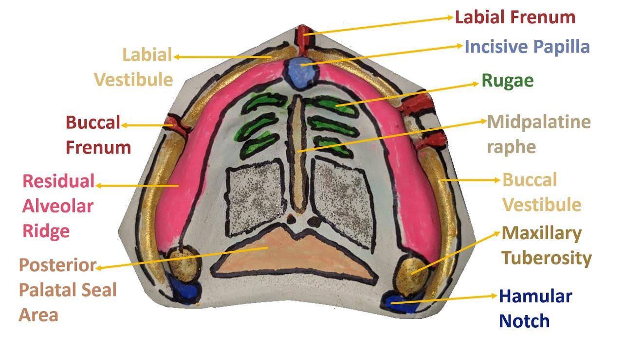
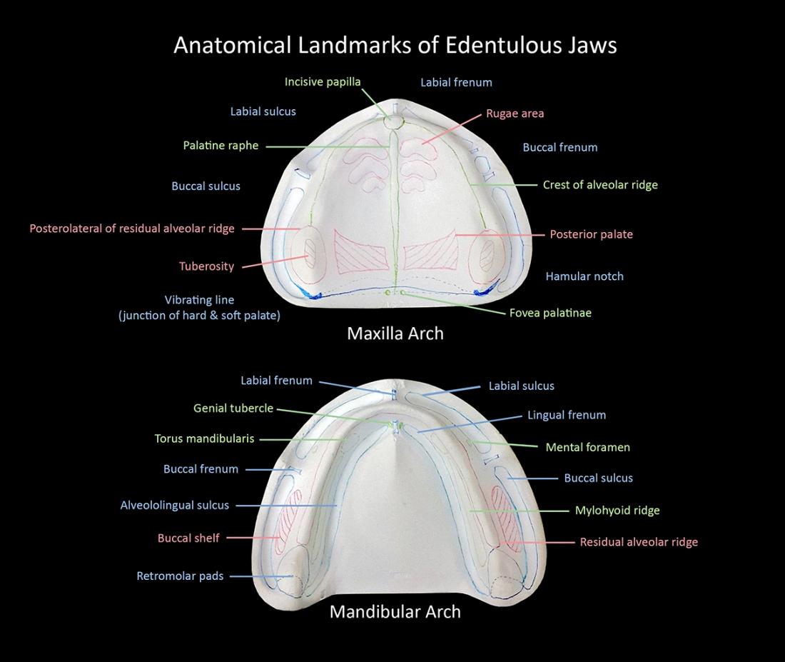
The hamular notch is essentially a narrow cleft of loose connective tissue between the distal surface of the maxillary tuberosity and the hamular process of the medial pterygoid plate. This structure serves as the posterior boundary of the maxillary denture, and it plays a crucial role in achieving a proper posterior palatal seal. In dental prosthetics, a full maxillary denture base is extended over the tuberosity and rests on the mucosa overlying the hamular notch, which is the area between the hamulus and the maxillary tuberosity.
So why is the hamular notch so important? For starters, it helps to ensure that the maxillary denture is securely in place. By extending the denture base over the tuberosity and resting it on the mucosa overlying the hamular notch, the denture is less likely to shift or move around in the patient’s mouth. This is important not just for comfort, but also for proper function – a denture that fits poorly can make it difficult to eat, speak, and carry out other everyday activities.
Additionally, the hamular notch plays a role in achieving a proper posterior palatal seal. This is the seal that is created between the maxillary denture and the patient’s palate, which helps to create suction and hold the denture in place. Without a proper posterior palatal seal, the denture is more likely to move around and cuse discomfort.
The hamular notch may be small, but it plays a vital role in dental prosthetics. By extending the denture base over the maxillary tuberosity and resting it on the mucosa overlying the hamular notch, a proper posterior palatal seal can be achieved and the denture can be held securely in place. So the next time you’re working on a maxillary denture, be sure to pay close attention to this important anatomical feature!
Understanding the Hamular Notch
The hamular notch is a small, V-shaped indentation located at the junction of the maxilla bone and the hamular process of the sphenoid bone. The hamular process is a small, hook-shaped projection that extends from the sphenoid bone and lies just beyond the distal end of the alveolar process of the maxilla. The hamular notch is an important anatomical landmark in the oral cavity, as it serves as a reference point for various dental and surgical procedures.
The hamular notch is located on the posterior border of the hard palate, approximately 1 cm lateral to the midline. It is bounded by the hamular process superiorly and the maxillary tuberosity inferiorly. The pterygoid hamulus, a small bony projection located just anterior to the hamular process, may also be palpable in this area.
In dentistry, the hamular notch is important for the placement of certain dental appliances, such as partial dentures and palatal expanders. It is also a reference point for local anesthesia injections and surgical procedures such as the removal of impacted thid molars. Additionally, the hamular notch plays a role in speech production and resonance, as it is a point of attachment for certain muscles involved in velopharyngeal closure.
The hamular notch is a small, V-shaped indentation located at the junction of the maxilla and the hamular process of the sphenoid bone. It serves as a reference point for various dental and surgical procedures, and plays a role in speech production and resonance.

Understanding the Hamular Notch in the Maxilla
The hamular notch, also known as the pterygo-maxillary notch, is a narrow cleft of loose connective tissue located between the distal surface of the maxillary tuberosity and the hamular process of the medial pterygoid plate. It is an important anatomical landmark that serves as the posterior boundary of the maxillary denture.
The hamular notch plays a crucial role in the construction of a well-fitting denture. Dentists use the hamular notch to achieve a posterior palatal seal, whih is the contact between the denture and the mucosa of the palate that prevents the entry of air and food particles into the oral cavity during swallowing. A properly sealed denture ensures maximum comfort and stability for the patient.
It’s important to note that the hamular notch is not always visible in all individuals. In some cases, it may be covered by a thick layer of soft tissue, making it difficult to locate. Dentists may use special techniques or instruments to find the hamular notch, such as palpation or imaging techniques like X-rays or CT scans.
The hamular notch is a small but important anatomical structure in the maxilla that helps dentists achieve a proper posterior palatal seal for denture wearers.
Location of the Hamular Notch
The hamular notch is an anatomical region located in the maxillary tuberosity area of the human skull. It is a small depression or notch located beteen the hamulus, a bony protrusion of the medial pterygoid plate of the sphenoid bone, and the maxillary tuberosity, which is the rounded prominence on the posterior aspect of the maxilla.
The hamular notch is an important landmark in dental prosthetic practice as a full maxillary denture base is extended over the tuberosity and rests on the mucosa overlying the hamular notch. This area is also known as the pterygomaxillary notch or the pterygoid notch.
In addition to its significance in dental prosthetics, the hamular notch is also an important anatomical landmark for maxillofacial surgery, as it serves as a reference point for various surgical procedures involving the maxilla and surrounding structures.
To summarize, the hamular notch is a small depression located between the hamulus and the maxillary tuberosity, which is an important landmark in dental prosthetics and maxillofacial surgery.
The Function of the Hamular Process
The hamular process, also known as the pterygoid hamulus, is a small bony structure that extends from the medial pterygoid plate of the sphenoid bone. The medial pterygoid plate is a thin, flat bone that forms part of the lateral wall of the nasal cavity and the medial wall of the pterygopalatine fossa.
The hamular process is a small, finger-like projection that extends downward and forward from the medial pterygoid plate. It is made of dense, compact bone and is located at the posterior end of the medial pterygoid plate.
The hamular process has several important functions in the human body. It serves as an attachment point for several muscles, including the tensor veli palatini muscle, the levator veli palatini muscle, and the superior constrictor muscle of the pharynx. These muscles play important roles in the functions of the soft palate, the pharynx, and the Eustachian tube.
One of the most important functions of the hamular process is to provide support for the soft palate. The soft palate is a muscular structure that separates the nasopharynx from the oropharynx and helps to prevent food and liquids from entering the nasal cavity during swallowing. The hamular process serves as an anchor point for the tensor veli palatini muscle, wich helps to tense the soft palate and keep it in position during swallowing.
The hamular process is a small but important structure in the human body. Its location and function make it an essential component of the soft palate and the muscles that control it.
The Location of the Hamular Process
The hamular process, also known as the pterygoid hamulus, is a bony hook-shaped process located bilaterally on each medial pterygoid plate of the sphenoid bone. It is positioned posterior and medial to each maxillary tuberosity. The pterygoid plates are two thin, flattened bony structures that arise from the sphenoid bone and extend dowward and forward to form the lateral walls of the nasal cavity. The medial pterygoid plate is located on the inner side of each pterygoid process and serves as an attachment site for several muscles, including the tensor veli palatini, levator veli palatini, and superior pharyngeal constrictor. The hamular process protrudes from the medial pterygoid plate and is an important landmark in the oral cavity as it serves as an attachment site for the palatopharyngeus muscle and the pterygomandibular raphe. the hamular process is located on the medial pterygoid plate of the sphenoid bone, posterior and medial to the maxillary tuberosity, and serves as an attachment site for important muscles in the oral cavity.

Source: youtube.com
The Importance of Hamular Notch
The hamular notch is a crucial area in achieving an effective posterior palatal seal in denture fabrication. It refers to the soft tissue region located between the distal surface of the tuberosity and the hamular process of the medial pterygoid plate. This notch plays a vital role in providing stability and retention to the denture.
When the denture extends too far into the hamular notch, it can cause irritation and soreness. On the oter hand, if the denture does not extend enough, it can result in poor retention, leading to instability and discomfort for the patient.
In dental practice, achieving the appropriate extension of the denture in the hamular notch area is critical to ensure the denture’s proper fit, stability, and retention. Dentists and dental technicians use various techniques to determine the optimal extension of the denture in this area, such as palpation, visual inspection, and radiographic analysis.
To summarize, the hamular notch is an essential area in denture fabrication, and its proper extension is crucial in achieving an effective posterior palatal seal, which provides stability and retention to the denture.
Formation of the Masseteric Notch
The masseteric notch is an anatomical structure located at the distobuccal corner of the retromolar area. It is formed due to the contraction of the masseter muscle, whch is one of the strongest muscles in the human body responsible for chewing and grinding food.
The masseter muscle is attached to the zygomatic arch and the mandible, and when it contracts, it pulls the mandible upwards and backwards, causing a depression or notch to form in the tissue at the distobuccal corner of the retromolar area.
It is important to note that when the mouth is opened widely, the borders in this area can cut into the tissue. Therefore, it is recommended to record the masseteric notch with the mouth slightly opened to avoid any discomfort or injury.
The masseteric notch is formed due to the contraction of the masseter muscle, and it is located at the distobuccal corner of the retromolar area.
The Post Dam Area: An Overview
The post dam area is a term used in dentistry to refer to the region located between the anterior and posterior vibrating lines. More specifically, it is the area that extends medially from one tuberosity to the other. In simpler terms, it is the space at the back of the mouth where the upper jaw meets the throat.
The term “vibrating lines” refers to two specific lines in the mouth: the anterior and posterior vibrating lines. The anterior vibrating line is a ridge located on the hard palate, while the posterior vibrating line is a ridge located on the soft palate.
The post dam area is an important region in dentistry because it is where the denture base will be plced in an upper denture. The denture base is the part of the denture that sits on the gums and supports the artificial teeth. By fitting the denture base in the post dam area, it ensures the denture is stable and comfortable for the patient.
It is also worth noting that the post dam area can vary in size and shape from person to person. Dentists must take this into account when designing and fitting dentures to ensure a proper fit. Factors that can affect the size and shape of the post dam area include age, gender, and the presence of any underlying health conditions.
To summarize, the post dam area is the region between the anterior and posterior vibrating lines, extending medially from one tuberosity to the other. It is a crucial area in dentistry, particularly when it comes to fitting and designing dentures. Dentists must consider the size and shape of the post dam area when creating a custom denture to ensure a proper fit and optimal comfort for the patient.
The Significance of the Primary Stress Bearing Area
The buccal shelf is referred to as the primary stress-bearing area in the mandibular arch because it is the region that bears most of the occlusal forces during mastication. When a person chews food, the force is transmitted through the teeth and onto the surrounding bone. The buccal shelf is located on the outer surface of the mandible and is parallel to the occlusal plane. This orientation makes it an ideal area for absorbing and distributing the stresses generated during mastication.
Moreover, the bone in the buccal shelf is dense and compact, which makes it more resistant to resorption than other areas of the mandible. This means that the buccal shelf can maintain its structural integrity and provde support to the teeth even in cases of bone loss or periodontal disease.
The buccal shelf is called the primary stress-bearing area because it is the region in the mandibular arch that is most capable of withstanding and distributing the occlusal forces generated during mastication. Its parallel orientation to the occlusal plane and dense bone structure make it a reliable area for supporting the teeth and maintaining stability in the mandibular arch.

Source: mydentaltechnologynotes.wordpress.com
The Location of the Vibrating Line
The vibrating line is a significant anatomical landmark in the oral cavity. It is an imaginary line that extends from one hamular notch to the other, passing approximately 2 mm ahead of the fovea palatini. This line marks the beginning of the movement in the soft palate when an individual pronounces the sound “ah”. In other words, it is the point where the soft palate starts to vibrate, causing the air to flow thrugh the oral and nasal cavities. The vibrating line plays a crucial role in the proper functioning of the oral cavity, particularly in speech and swallowing. It helps to separate the oral and nasal cavities, allowing us to produce various sounds and prevent food and liquid from entering the nasal cavity during swallowing.
The Meaning of Buccal Shelf Area
The buccal shelf area can be defined as the bony structure that is located bilaterally on the posterior lateral area of the mandible. It is an important anatomical landmark in dentistry, particularly in the placement of dentures and implants.
More specifically, it is bounded medially by the alveolar bone, which is the bone that supports the teeth. Laterally, it is bounded by the external oblique ridge, which is a bony ridge on the outside of the mandible. Anteriorly, it is bounded by the buccal frenum, which is a fold of tissue that attaches the cheek to the gum. it is bounded posteriorly by the retromolar pad, which is a soft tissue pad located behid the last molar in the mandible.
The buccal shelf area plays an important role in providing support and stability for dentures and implants. It is also important in determining the height and width of the mandible, which can impact the overall appearance of the face. In addition, the buccal shelf area can be used as a reference point for radiographic imaging and surgical procedures.
The buccal shelf area is a critical anatomical structure in dentistry, serving as a key reference point for a variety of dental procedures. Its location and boundaries are defined by the alveolar bone, external oblique ridge, buccal frenum, and retromolar pad.
Attachment of the Hamular Process
The hamular process, a small hook-shaped projection of bone located at the posterior end of the medial pterygoid plate of the sphenoid bone, serves as an attachment site for several structures in the oral and pharyngeal regions.
One of the structures that attaches to the hamular process is the tensor veli palatini muscle. This muscle originates from the scaphoid fossa, the spine of the palatal aponeurosis, and the lateral wall of the cartilaginous auditory tube. The muscle then winds its tendon around the hamular process in a groove before inserting into the soft palate and the transverse bony ridge on the posterior border of the hard palate.
Other structures that attach to the hamular process include the levator veli palatini muscle, which originates from the temporal bone and inserts into the soft palate, and the superior constrictor muscle of the pharynx, which attaches to the posterior border of the hamulus.
The hamular process plays an important role in the anatomy and function of the oral and pharyngeal regions by providing attachment sites for several important muscles and othr structures.
Attachment of the Hamulus
The hamulus is a small hook-shaped bony process located on the medial pterygoid plate of the sphenoid bone. It serves as an attachment point for several structures, including the tensor tympani and tensor veli palatini muscles.
The tensor tympani muscle originates from the cartilaginous portion of the auditory tube and the adjacent bone, and its tendon passes through the canal above the cochlear window before attaching to the malleus bone of the middle ear. When this muscle contracts, it pulls the malleus medially, threby reducing the amplitude of sound vibrations transmitted through the ossicular chain.
The tensor veli palatini muscle originates from the scaphoid fossa of the sphenoid bone and the cartilage of the auditory tube, and its fibers converge to form a tendon that loops around the hamulus before inserting into the palatal aponeurosis. When this muscle contracts, it pulls the soft palate and the aponeurosis medially and superiorly, thereby helping to open the auditory tube and equalize air pressure between the middle ear and the nasopharynx.
The hamulus serves as a common attachment point for the tensor tympani and tensor veli palatini muscles, which are important for hearing and pressure regulation in the middle ear and nasopharynx, respectively.

The Role of the Pterygoid Muscle
Pterygoid is an adjective used in anatomy to describe something that is relaed to or located in the inferior part of the sphenoid bone of the vertebrate skull. The sphenoid bone is a complex bone that is located in the middle of the skull and is involved in the formation of the base of the skull and the orbits, which house the eyes.
The pterygoid region is located in the posterior part of the skull and is made up of the pterygoid plates, which are two thin, flat bones that extend from the inferior surface of the sphenoid bone. The pterygoid muscles, which are responsible for movements of the jaw, are also located in this region.
The pterygoid muscles are divided into two groups: the lateral pterygoid muscles and the medial pterygoid muscles. The lateral pterygoid muscles are involved in opening the mouth and moving the jaw from side to side, while the medial pterygoid muscles are involved in closing the mouth and elevating the jaw.
Pterygoid is a term used in anatomy to describe the region and structures located in the inferior part of the sphenoid bone of the skull, including the pterygoid plates and the pterygoid muscles, which are important for jaw movements.
Conclusion
The hamular notch, also known as the pterygo-maxillary notch, is an important anatomical structure located at the junction of the maxilla and the hamular process of the sphenoid bone. It is a narrow cleft of loose connective tissue that serves as the posterior boundary of the maxillary denture and aids in achieving a posterior palatal seal.
In dental prosthetic practice, a full maxillary denture base is extended over the tuberosity and rests on mucosa overlying the hamular notch. This area between the hamulus and the maxillary tuberosity is crucial for the stability and retention of the denture, as it provides a secure posterior stop for the denture and prevents it from dislodging durig function.
The hamular process, from which the hamular notch derives its name, is a small projection of bone that extends inferiorly from the medial pterygoid plate. It serves as an attachment site for the tensor veli palatini muscle, which plays a crucial role in the opening and closing of the eustachian tube.
The hamular notch is an important anatomical structure in dental prosthetic practice that provides stability and retention for the maxillary denture. Its location at the junction of the maxilla and hamular process makes it a crucial posterior boundary for the denture, and its relationship to the hamular process highlights its role in the opening and closing of the eustachian tube.
