Cranial bones are an essential part of the human anatomy, providing protection for the brain and providing structural support for the face. But how do cranial bones develop? In this blog post, we’ll explore the process of cranial bone development and discuss why it is so important to our overall health.
Cranial bones are formed through intramembranous ossification, which occurs when a fibrous membrane is formed and is later replaced with bone tissue. Intramembranous ossification directly converts the mesenchymal tissue to bone and forms the flat bones of the skull, clavicle, and most of the cranial bones. This development begins by the end of the first month as a condensation and thickening of mesenchymal tissue surrounding the head end of the notochord (a type of embryonic tissue).
The process of intramembranous ossification is vital to proper cranial bone formation as it helps ensure that our heads develop properly into adulthood. Without intramembranous ossification, our skulls may not be able to provide enugh structural support or protection for our brains. Additionally, intramembranous ossification helps form facial features such as cheekbones, brows, and chin that give us our individual identity.
It is important to note that proper cranial bone development does not occur in isolation; other factors such as nutrition and environment also play a role in proper cranium growth. For example, nutrient deficiencies can lead to underdevelopment or malformation in cranial bones which can lead to physical deformities or medical conditions such as craniosynostosis (premature fusion of skull sutures). Therefore, it is essential for parents to take steps to ensure their children have access to adequate nutrition during their early stages of development in order to maximize their chances for healthy skull growth.
In conclusion, understanding how cranial bones develop is important for maintaining good health. Proper intramembranous ossification helps form our skulls into adulthood while factors such as nutrition and environment also play a role in our overall skull health. By taking steps to ensure children have access to adequate nutrition during early development parents can help maximize their children’s chances for healthy skull growth.
Development of Cranial Bones
Cranial bones develop from mesenchymal tissue located around the head end of the notochord. This process begins during the first month of development when the mesenchyme thickens and condenses into distinct masses, which are then organized and shaped into the elements that make up the cranial bones. As development continues, these elements grow and fuse together to form a fully functional cranial skeleton.

Source: wellnesslifezone.com
Development of Cranial Bones
Cranial bones develop through a process known as endochondral ossification. This is a two-stage process that begins with the formation of cartilage and ends with the replacement of cartilage with bone tissue. During the first stage, a matrix of connective tissue forms around the cartilage model, which prvides nutrients to the growing cells. As these cells enlarge and divide, they form layers of new cartilage. This process is known as chondrification.
In the second stage of endochondral ossification, some of these chondrocytes become osteoblasts, which are cells that secrete calcium salts and proteins to form bone tissue. The osteoblasts replace the cartilage until all that remains is a bony structure that has replaced the original cartilage model. This is how cranial bones develop quizlet. Over time, this bony structure matures and becomes stronger as it is remodeled by other types of cells such as osteoclasts and osteocytes.
Development of Cranial Bones Within the Osseous Membrane
Yes, cranial bones do develop within the osseous membrane. This is known as intramembranous ossification, which happens when a fibrous membrane is formed and later replaced with bone tissue. The process begins with the formation of mesenchymal tissue that develops into a membranous structure. This structure then gives rise to immature bone cells, which later develop into mature bone cells. As these cells mature, they gradually lay down new layers of bone matrix and become organized into distinct structures that make up the cranial bones.
Formation of Cranial Bones Through Ossification
Intramembranous ossification is the process by whch certain bones of the skull, such as the flat bones of the skull, clavicle, and most of the cranial bones, form directly from mesenchymal tissue. This process involves the transformation of mesenchymal tissue into bone tissue without a cartilage template or intermediary. The mesenchymal cells condense and differentiate into osteoblasts (bone-forming cells), which then begin to secrete collagen fibers and make mineral deposits in order to form bone. Intramembranous ossification is a gradual process that occurs in several stages, from formation of a primary center of ossification to secondary centers and finally to full maturation. As intramembranous ossification progresses, new bone is formed from existing bone material and remodeled over time to its final shape.
Fusing of Cranial Bones in Adolescence
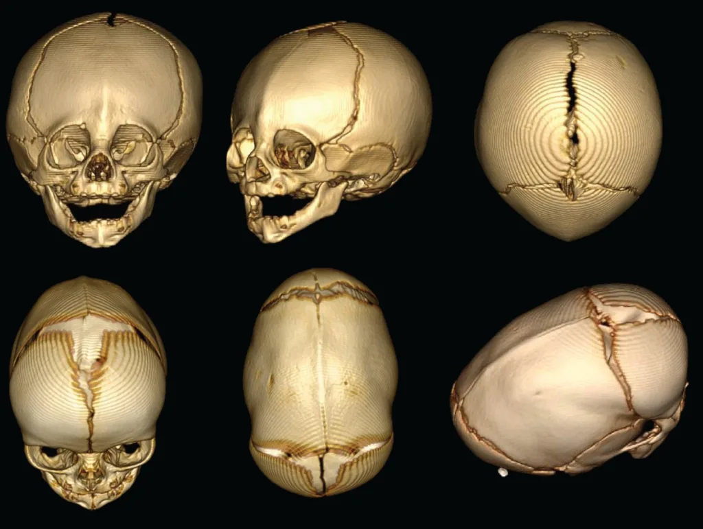
At approximately two years of age, the cranial bones of a typical child begin to fuse together. This process is known as craniosynostosis and occurs when the sutures that separate the cranial bones become filled with calcium and turn into bone. During this process, the shape of the head can change as some of the sutures close earlier than others, resulting in an abnormal head shape or size. If left untreated, craniosynostosis can cause increased pressure inside the skull which can lead to neurological problems. Treatment for this condition usually involves early diagnosis and surgery to correct any abnormalities.

Development of the Skull at Birth
No, a baby’s skull is not completely developed at birth. The bones in the skull are soft and flexible, allowing them to move and overlap during childbirth. This helps the baby pass through the birth canal without causing any damage. After birth, it takes 9-18 months for the bones of the skull to fully harden and fuse together. During this time, some babies may develop positional plagiocephaly, a condition where the shape of their head becomes misshapen due to laying in one position for too long.
Development and Growth of Bones
The development and growth of bones is a complex process known as osteogenesis or ossification which involves the transformation of progenitor cells into mature bone cells. It is a vital part of skeletal development that starts in the womb and continues throughot life. This process involves three stages: proliferation, maturation of matrix, and mineralization. During proliferation, progenitor cells divide to form osteoblasts, which are responsible for the formation of bone tissue. During maturation of matrix, these cells produce proteins such as collagen that form a scaffold on which calcium and other minerals are deposited to create strong bones. Finally, during mineralization, calcium salts become embedded in the matrix to make it even stronger. With healthy eating and regular exercise, bones can stay strong throughout life by constantly replenishing the minerals necessary for their growth and maintenance.
Development of Bones in a Fetus
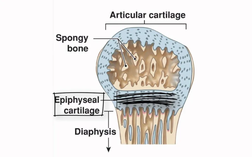
During fetal development, bones form through a process known as endochondral ossification. This process begins when a cartilaginous template, or scaffold, is formed from mesenchymal cells in the embryo. This cartilage is then progressively replaced with bone tissue, which is laid down in layers by specialized cells called osteoblasts. As these layers of bone grow and thicken, the cartilage will eventually be completely replaced unil the bone reaches its full size. The process of endochondral ossification continues throughout childhood and adolescence until growth plates located at either end of long bones fuse during puberty. At this point, growth ceases and the bones have reached their adult size and shape.
Bones Developed Through Intramembranous Ossification
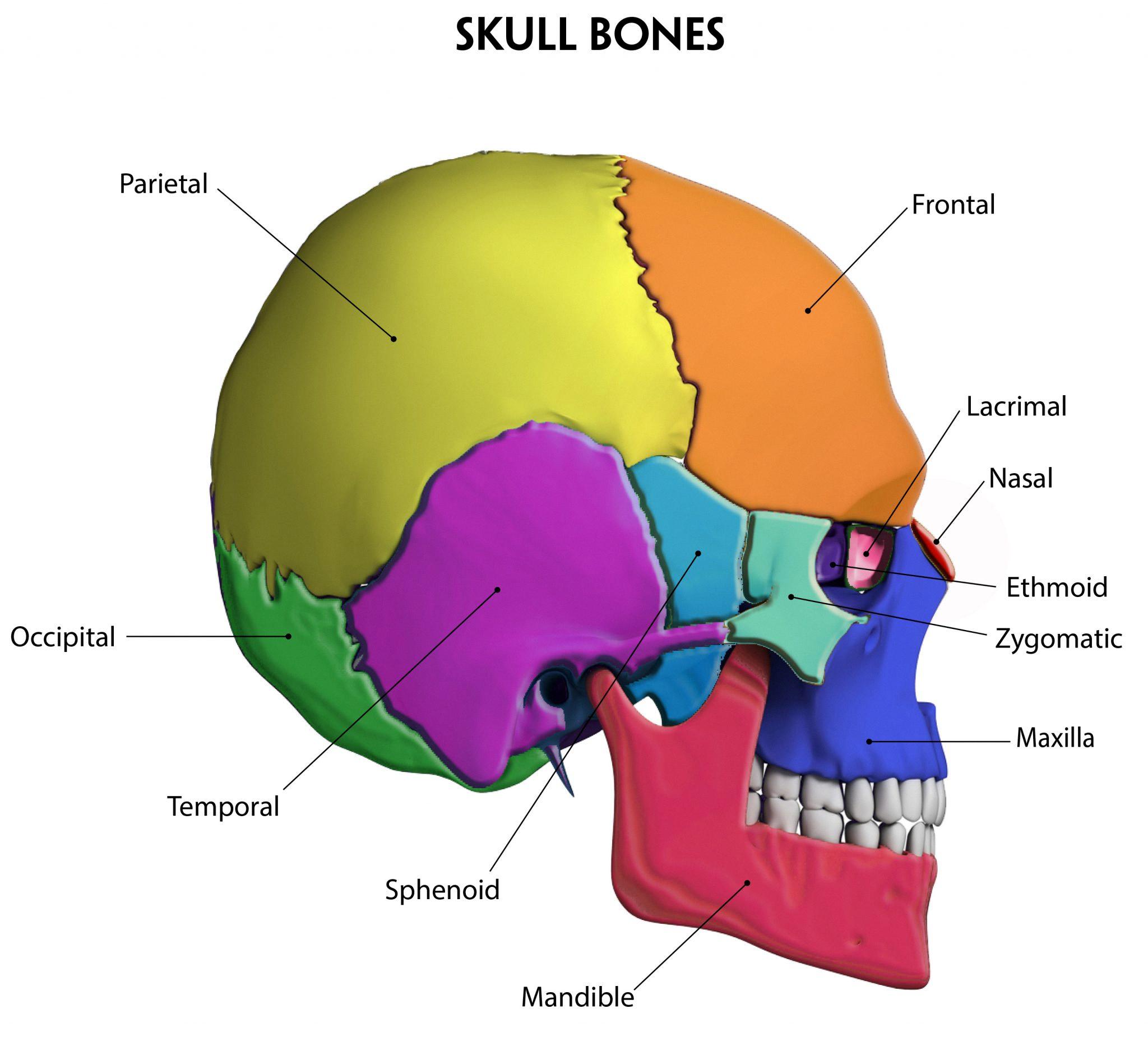
Intramembranous ossification is the process of bone formation from fibrous membranes, and is responsible for the development of several bones in the body. The flat bones of the skull, such as the frontal, parietal and temporal bones, as well as the mandible and clavicles, all form via intramembranous ossification. In addition to these bones, some areas of certain othr bones form via intramembranous ossification. This includes parts of the sphenoid and ethmoid bones in the skull, as well as parts of the maxilla and zygomatic arch. Intramembranous ossification also contributes to some joints (such as those between two adjacent flat bones) and help to form sutures that connect skull plates together.

Does Mesoderm Contribute to Bone Development?
Yes, bone development does occur from mesoderm. Mesoderm is one of the three primary germ layers formed in the early stages of embryonic development. In most cases, bone formation begins when mesenchymal cells differentiate into osteoblasts, which then secrete proteins to form a collagen matrix. This collagen matrix is then mineralized and hardened to give rise to the bones. The bones in the axial skeleton mostly derive from the mesoderm layer, except for some bones in the skull which come from the ectoderm. All the bones in the appendicular skeleton also derive from mesoderm. Bone formation is an essential part of skeletal development, and without it our bodies wold not be able to provide us with structure or movement.
Development of the Skull Bone from Axial Mesoderm
The axial mesoderm is responsible for the formation of the intraparietal portion of the occipital bone and the parietal bone. Both of these bones are formed through a process called intramembranous ossification, which involves the deposition of calcium salts to form hard, bony tissue. The occipital bone consists of four parts: the squamous portion, two lateral portions (the temporal and parietal) and a basal part (the occipital condyle). The parietal bone consists of two plates that meet at the coronal suture. Together, these bones form much of the lateral sides and roof of the skull.
Development of Bones Within a Fibrous Membrane
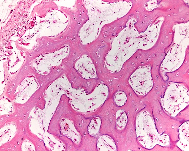
The bones that develop within a fibrous membrane through intramembranous ossification are the flat bones of the skull, such as the frontal, parietal, temporal, sphenoid and occipital bones; the mandible or lower jawbone; and the clavicles or collarbones. During this process, mesenchymal cells form a template of the future bone and differentiate into osteoblasts which deposit calcium phosphate to form the hard matrix of bone tissue. This gradually thickens to produce a compact layer beteen two layers of connective tissue. As ossification progresses, cartilage is replaced by bone tissue and spongy bone forms in the interior sections. The process is complete with a well-formed trabecular pattern of cancellous bone covered by a thin sheet of cortical bone on its exterior surface.
Endochondral Ossification and the Formation of the Cranial Base
Yes, the cranial base is formed by endochondral ossification. This process begins with the formation of a cartilage primordium from condensed mesenchymal cells. This is then followed by chondrocyte proliferation which maintains the synchondroses and leads to the elongation of the cranial base. The endochondral ossification process is important for the formation of bones as it involves a transition from cartilage to bone tissue. This transition begins with the calcification of cartilage, resulting in its gradual dissolution and replacement with bone tissue. The result is an increase in strength and stability of the structure.

Endochondral Ossification and the Formation of Bones
Endochondral ossification is a process which involves the transformation of cartilage into bone. It is responsible for the formation of all long bones in the human body – including those in the axial skeleton (vertebrae, ribs) and appendicular skeleton (limbs). Specifically, this includes bones such as the femur, humerus, radius and ulna, tibia and fibula, metacarpals, phalanges and carpals. Additionally, it is involved in the formation of some facial bones such as the mandible and maxilla. During endochondral ossification, a cartilaginous model of the bone forms first before it is gradually replaced by bone tissue. This process generally begins in utero but can continue up until adolescence as bones continue to grow and develop.
Endochondral Ossification and the Bones of the Skull
The bones of the skull that form by endochondral ossification are the temporal, sphenoid, zygomatic, and basioccipital bones. These bones are all part of the cranial base and form through a process known as endochondral ossification. This process involves the transformation of cartilage into bone tissue. It begins with the formation of a cartilaginous template around which bone is gradually deposited. As this occurs, the template is gradually replaced by bone until it has been fully replaced. The result is a strong, rigid bone structure that forms a key part of the skull’s structure and protection for the brain.
Conclusion
In conclusion, cranial bones are formed through intramembranous ossification, which occurs when a fibrous membrane is formed and is later replaced with bone tissue. This process begins in the mesenchymal tissue surrounding the head end of the notochord and occurs within fibrous connective tissue membranes. Endochondral ossification also leads to the formation of clavicles and some of the cranial bones. Together, these processes enable the development of a fully formed skull, which is essential for protecting the delicate brain and providing structure to our heads.
